
Lyophilization Explained: Why Research Peptides Ship as Powder

Research-only note: This article is for educational purposes and discusses a compound intended strictly for in vitro and laboratory research. The information below is not medical advice, and the products referenced are not for human consumption.

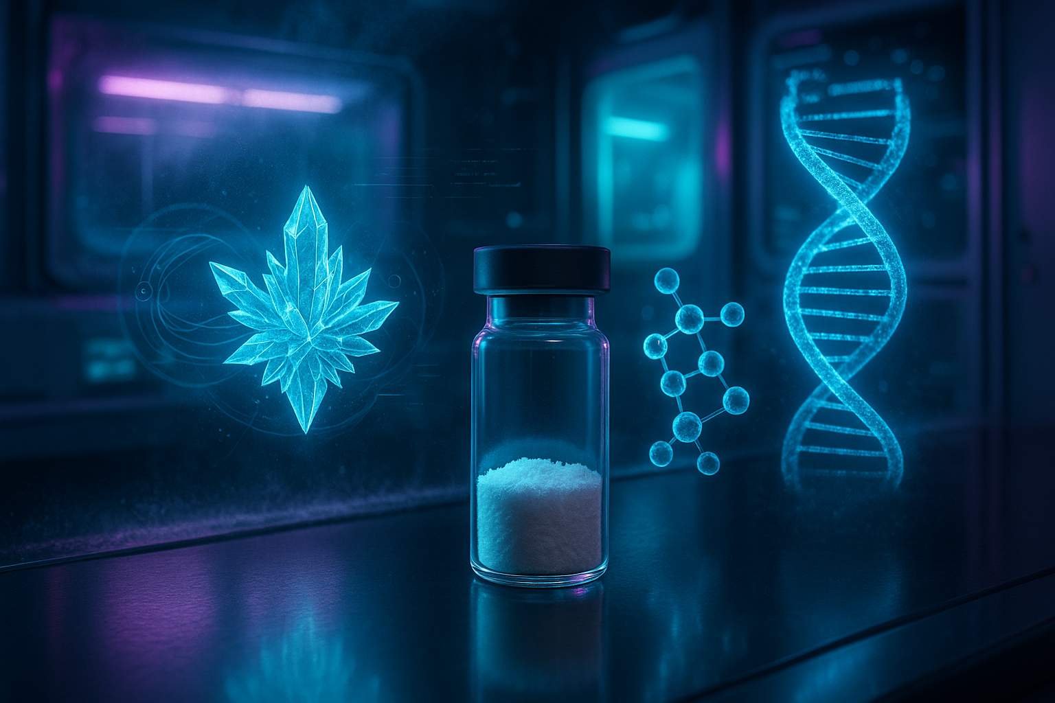
Peptide lyophilization is the controlled freeze-drying process that turns an aqueous peptide solution into a dry, porous solid by removing water through sublimation under vacuum rather than by evaporation. It is why a vial of Ipamorelin 10mg arrives as a white cake instead of a liquid: in the dry amorphous state the compound is far more stable, tolerates ambient shipping, and holds its purity specification for years rather than days. Understanding peptide lyophilization is the fastest way to judge whether a vial has travelled well.

Key takeaways

  • Peptide lyophilization removes water by freezing the solution and subliming the ice under deep vacuum, so the compound never passes through a destabilising warm liquid phase.
  • The cycle has three defined stages — freezing, primary drying and secondary drying — and each is a separate control point.
  • Water is the main reactant in peptide degradation; removing it suppresses hydrolysis, deamidation and aggregation.
  • The visible cake is a process record: an intact, uniform cake indicates drying stayed below the collapse temperature.
  • Residual moisture is driven below 1–2%, because leftover water plasticises the amorphous glass and restarts degradation chemistry.
  • The powder peptide lyophilization produces is a storage format, not a finished preparation — it still has to be reconstituted correctly.

What is peptide lyophilization?

Freeze-drying exploits the phase behaviour of water below its triple point. Drop the pressure below roughly 6 mbar and ice no longer melts on warming — it converts straight to vapour. Peptide lyophilization uses that shortcut deliberately: the solution is frozen solid first, then water is pulled out as vapour, so the peptide never sees a concentrated, warm liquid environment. Most of the damage in conventional drying happens in exactly that phase.

The output is an amorphous glass rather than a crystal. Molecular mobility in that glass is orders of magnitude lower than in solution, and reactive partners are not mobile enough to meet. Peptide lyophilization is the dominant solidification route in pharmaceutical development for this reason, and the process trade-offs are reviewed in the pharmaceutical freeze-drying literature.

  • Physical form: an amorphous, highly porous glass with large internal surface area.
  • Water content: reduced to low single-digit percentages, often below 1%.
  • Reconstitution behaviour: the porous structure rewets in seconds, which is why the cake dissolves fast.
  • Practical result: ambient shipping becomes viable without a validated cold chain.

How the peptide lyophilization cycle works

A peptide lyophilization run is a sequence of three engineered stages, each with its own failure mode. Vials are filled with peptide solution — often with a bulking agent such as mannitol or a stabilising sugar such as trehalose — and partially stoppered before cooling.

  • Freezing: shelf temperature drops to roughly ?40 °C, converting bulk water to ice and concentrating the peptide between the crystals.
  • Ice nucleation control: freezing rate sets crystal size, which sets pore size in the cake and therefore the drying rate.
  • Primary drying: vacuum is applied and heat supplied just fast enough to sublime the ice — the longest phase, removing most of the water.
  • Collapse-temperature limit: product temperature must stay below the collapse temperature, a few degrees above the glass transition of the concentrate.
  • Secondary drying: shelf temperature is raised to desorb the bound water remaining after the ice is gone.
  • Moisture endpoint: peptide lyophilization cycles end on a defined residual-moisture target, not on clock time.
  • Stoppering: vials are sealed under vacuum or backfilled with inert gas before the chamber opens.

The collapse limit dominates cycle design because sublimation is itself a cooling process. Push heat in too hard and the frozen matrix softens and slumps before the ice is gone. Everything else in peptide lyophilization sits downstream of holding that line.

Peptide lyophilization versus solution storage

The clearest case for the dried format compares the three states a research peptide can exist in. Solution-phase peptides degrade through water-mediated chemistry; dry peptides largely do not.

Format Dominant degradation route Practical shelf life Shipping tolerance
Lyophilized powder, sealed Minimal; limited by residual moisture Long-term at ?20 °C; months at ambient High — survives ambient transit
Aqueous solution, refrigerated Hydrolysis, deamidation, oxidation, adsorption to glass Days to a few weeks Low — requires cold chain
Aqueous solution, frozen aliquots Freeze–thaw stress and aggregation Weeks to months if never re-thawed Very low — thaw excursions unrecoverable
  • Hydrolysis needs water: backbone amide bonds cannot be cleaved hydrolytically when almost no water is present.
  • Deamidation is moisture-dependent: asparagine and glutamine residues are far more reactive in solution than in a dry glass.
  • Oxidation targets methionine and tryptophan: dissolved oxygen and trace metals accelerate it, and both are reduced in a dry sealed vial.
  • Adsorption losses disappear: dilute peptides lose material to container surfaces, which peptide lyophilization defers until reconstitution.
  • Dilution is deferred: the researcher picks the final concentration, so one vial suits different assay designs.

Research applications of the dried format

Nearly every peptide used as a laboratory tool compound ships dry. The rationale appears throughout the literature on parenteral peptide and protein delivery, where peptide lyophilization is the default when a molecule is only marginally stable in water.

  • Reference standards: a dry, mass-defined vial is the cleanest basis for calibration curves.
  • Binding and signalling assays: researchers reconstitute to an exact stock molarity rather than trusting a shipped concentration.
  • Long-duration designs: studies running for months can draw from one validated lot.
  • Multi-site work: identical dry vials ship without every site needing cold-chain logistics.
  • Blends: co-lyophilised products such as the GLOW Blend (GHK-Cu, TB-500, BPC-157) hold a fixed component ratio that a solution could not.
  • Comparative pharmacology: tool compounds such as Tesamorelin 10mg and Ipamorelin are studied side by side from equivalently characterised dry material.

Reading the cake: what powder appearance signals

Because peptide lyophilization writes its own process record into the solid, cake appearance is diagnostic. Inspect a vial before reconstituting it — and note that peptide lyophilization at low fill volumes can legitimately produce something that looks like nothing at all.

  • Intact white cake: the expected result — the structure held and drying was uniform.
  • Thin film or scattered flakes: normal at low fill volumes; 10 mg is a very small mass across a vial base.
  • Shrunken or retracted cake: the matrix pulled from the glass, usually cosmetic, but worth the lab record.
  • Collapsed or glassy melt-back: the product exceeded its collapse temperature, which correlates with higher residual moisture.
  • Discolouration: yellowing is not a normal outcome and warrants querying the lot.
  • Visible moisture: a seal-integrity concern, since water ingress restarts hydrolytic chemistry.

These checks are cheap and immediate. Pair them with lot documentation and a disciplined peptide storage routine.

Handling, reconstitution, and quality verification

  • Equilibrate before opening: let a cold vial reach room temperature so moisture does not condense onto the cake.
  • Store sealed and dark: unopened vials are typically held at ?20 °C, protected from light and moisture.
  • Reconstitute gently: run the diluent down the vial wall rather than jetting it onto the cake, and swirl instead of shaking.
  • Choose the diluent deliberately: bacteriostatic or sterile water is standard for in vitro stock preparation, and the choice should be logged.
  • Aliquot immediately: in solution the stability clock restarts, so single-use aliquots prevent repeated freeze–thaw cycles.
  • Log the concentration: record diluent volume against labelled mass so stock molarity stays traceable.
  • Check the documentation: every NeuroPept Labs lot ships with batch-specific third-party analytics, and COA validity is confirmed at freedomdiagnosticstesting.com using the Accession Number, Client ID, or Search Code shown in the product images.
  • Follow a written protocol: the full sequence is covered in the guide to reconstituting research peptides.

Considerations for experimental design

  • Lot consistency: plan long studies around one lot, since peptide lyophilization parameters differ marginally between runs.
  • Excipient awareness: bulking agents and stabilisers are part of the vial contents and matter in sensitive cell systems.
  • Stock age: treat reconstituted stock age as a variable and record it with the result.
  • Temperature logging: document freezer excursions rather than assuming stability held.
  • Container effects: account for adsorption at low stock concentrations in plastic labware.
  • Purity floors: define a minimum acceptable HPLC purity before ordering material.

Clean data begins upstream of the assay. A sound peptide lyophilization cycle, an intact cake, verifiable third-party analytics and a disciplined reconstitution record together mean that variance at the bench belongs to the biology, not the material — the whole argument for sourcing documented compounds such as research-grade Ipamorelin from a supplier that publishes batch-level data.

Frequently asked questions

What is peptide lyophilization?

Peptide lyophilization is freeze-drying applied to peptides. The solution is frozen solid, then water is removed by sublimation under vacuum and by desorption at raised temperature, leaving a dry amorphous cake that is far more chemically stable than the original liquid.

Why are research peptides shipped as a lyophilized powder instead of a solution?

Water drives the main degradation pathways, including hydrolysis, deamidation and oxidation. Peptide lyophilization removes it, so a vial survives ambient shipping and long storage and the researcher chooses the final concentration.

What does a collapsed or shrunken cake indicate?

Shrinkage where the cake pulls away from the glass is often cosmetic. A genuinely collapsed cake suggests the product exceeded its collapse temperature during drying, which is associated with higher residual moisture and faster degradation, so the lot documentation should be reviewed.

Does the amount of visible powder show how much peptide is in the vial?

No. A 10 mg fill is a very small mass and can appear as a thin film or a few flakes after transit. Labelled mass is confirmed by the certificate of analysis, not by how full the vial looks.

How long does a lyophilized research peptide stay stable?

Sealed at around minus 20 degrees Celsius and protected from light and moisture, dry peptides are generally stable for long periods, while reconstituted solutions are measured in days to weeks. Residual moisture and temperature excursions are the limiting factors.

Are lyophilized research peptides approved for human use?

No. All peptides supplied by NeuroPept Labs are sold for in vitro and laboratory research only. They are not approved for human or veterinary use, are not intended to diagnose, treat, cure or prevent any disease, and are not for human consumption.

Research-use-only disclaimer: All products referenced are sold for laboratory and research use only. They are not intended to diagnose, treat, cure, or prevent any disease, and are not for human or veterinary consumption. Researchers sourcing documented, high-purity material can review Ipamorelin 10mg from NeuroPept Labs, supplied as a lyophilized research compound with batch-specific third-party analytics.

Peptide Half-Life Explained: What Determines Duration of Action

Research-only note: This article is for educational purposes and discusses a compound intended strictly for in vitro and laboratory research. The information below is not medical advice, and the products referenced are not for human consumption.

Peptide half-life is the time required for the concentration of a peptide in a biological matrix to fall by fifty percent. It is the number that best predicts how long a research compound stays measurable in a model system, and it is set almost entirely by two forces: enzymatic cleavage and renal filtration. Peptide half-life is also what explains why two structurally similar analogues such as CJC-1295 No DAC and its albumin-bound counterpart behave so differently in the same assay.

Key takeaways

  • Peptide half-life is governed by proteolytic degradation, glomerular filtration, and target-mediated uptake acting in parallel.
  • Dipeptidyl peptidase-4 (DPP-4) is the canonical cleavage route, snipping an N-terminal dipeptide whenever position two is alanine or proline.
  • Single-residue substitutions — D-alanine, Aib, glutamine, serine — block that cleavage and can extend peptide half-life several-fold.
  • Albumin binding, whether covalent (DAC) or non-covalent (fatty-acid acylation), moves duration from minutes into days.
  • Size matters: unmodified peptides sit far below the ~45–60 kDa glomerular cut-off and are filtered almost as fast as they appear.
  • Peptide half-life is a solution-phase property and says nothing about lyophilized shelf stability, which is a separate storage question.

What is peptide half-life?

In pharmacokinetic terms, peptide half-life (t½) describes first-order elimination: after one half-life half the compound is gone, after two half-lives three quarters. For small linear peptides the number is usually minutes rather than hours, because a short unprotected chain offers many attack points to peptidases while being small enough to pass the kidney’s filtration barrier unhindered.

Researchers also distinguish elimination half-life from the duration of measurable downstream effect: a growth hormone secretagogue may clear from plasma long before the IGF-1 response it triggered returns to baseline. Reporting peptide half-life without stating the compartment and the analyte measured is a common source of contradictory figures.

  • Plasma half-life — decay of the parent peptide itself, usually by LC-MS/MS.
  • Effect half-life — decay of a downstream marker; often much longer.
  • Terminal half-life — the final log-linear phase, which dominates dosing interval logic in a model.
  • Matrix dependence — serum, plasma with protease inhibitors, and buffer each give different answers.

Mechanism of action: how peptides are cleared

Three routes dominate. Proteolysis by exopeptidases and endopeptidases fragments the chain; glomerular filtration removes anything with a hydrodynamic radius below the renal cut-off; and receptor-mediated endocytosis internalises the compound alongside its target. The relative weight of each route is what a structural chemist manipulates when trying to lengthen peptide half-life.

DPP-4 (CD26) deserves particular attention. It is a serine exopeptidase that removes the first two residues from any chain presenting alanine or proline at position two — a motif shared by native GHRH, native GLP-1, and many endogenous signalling peptides. Published enzymatic work shows that replacing that single alanine measurably increases resistance to DPP-4, confirming the residue as the rate-limiting vulnerability rather than the chain as a whole.

  • DPP-4 cleavage — removes an N-terminal dipeptide; the single largest determinant of peptide half-life for GHRH- and incretin-class chains.
  • Neprilysin and other endopeptidases — cut internal bonds, producing multiple inactive fragments.
  • Aminopeptidases and carboxypeptidases — nibble from either terminus; C-terminal amidation blocks the latter.
  • Glomerular filtration — free peptides under roughly 45–60 kDa pass into urine; most research peptides are 1–5 kDa.
  • Target-mediated disposition — receptor binding and internalisation removes compound in a saturable, dose-dependent way.
  • Hepatic uptake — a secondary contributor for lipidated and larger conjugates.

Structural defences and how they compare

Medicinal chemistry answers each clearance route with a specific countermeasure. Proteolysis is blocked at the residue level; renal filtration is defeated by making the molecule effectively larger, usually by attaching it to serum albumin. The table below sets out literature-reported durations for well-characterised research compounds.

Compound (research context) Class Principal structural defence Reported half-life in literature
Native GHRH (1-44) Endogenous releasing hormone None — Ala at position 2 Minutes
CJC-1295 No DAC (Mod GRF 1-29) GHRH analogue Four substitutions including D-Ala2 Roughly half an hour
CJC-1295 with DAC GHRH analogue Covalent albumin conjugation via maleimide linker Approximately 6–8 days
Tesamorelin GHRH analogue N-terminal trans-3-hexenoyl cap Tens of minutes
Ipamorelin GHS-R pentapeptide D-amino acids, Aib, C-terminal amide Around two hours
Native GLP-1 Incretin None — Ala at position 8 1–2 minutes
Acylated incretin analogues Incretin analogue Aib substitution plus fatty-acid albumin binder Days

The pattern is consistent: residue-level edits move peptide half-life from minutes to tens of minutes, while albumin engagement moves it into days. Published pharmacokinetic work on the DAC-modified GHRH analogue estimated a half-life of 5.8 to 8.1 days — a roughly thousand-fold extension achieved with one conjugation chemistry (Teichman et al., J Clin Endocrinol Metab, 2006).

  • D-amino acid substitution — proteases are stereospecific for L-residues, so a single inversion blocks the cut.
  • Aib (?-aminoisobutyric acid) — steric bulk at position two makes the site unreadable to DPP-4.
  • N-terminal acylation — a hexenoyl or similar cap denies aminopeptidases a free terminus.
  • C-terminal amidation — neutralises the carboxylate and stops carboxypeptidase trimming.
  • Cyclisation and lactam bridges — conformational constraint hides scissile bonds.
  • Fatty-acid acylation — non-covalent albumin piggy-backing that is reversible and dose-tunable.
  • Covalent albumin conjugation (DAC) — an irreversible bond to Cys34 of serum albumin.
  • PEGylation — raises hydrodynamic radius above the renal cut-off without touching the sequence.

Research applications

Peptide half-life is not merely descriptive; it is an experimental variable in its own right, and a large body of PubMed-indexed literature is devoted to engineering it. Albumin-binding ligand work has demonstrated elimination half-life extensions of roughly twenty-five-fold in rodent models, illustrating how far the piggy-back strategy can be pushed (Zorzi et al., Nature Communications, 2017).

  • Serum-stability assays that quantify degradation rate in the presence of native protease activity.
  • Modelling pulsatile versus continuous receptor exposure in cell culture.
  • LC-MS/MS metabolite identification to map actual cleavage sites.
  • Albumin-binding affinity measurement by surface plasmon resonance.

Why peptide half-life shapes GH-axis study design

Nowhere is this more visible than in growth hormone secretagogue research. The somatotropic axis responds to pulses, not plateaus, so a long-acting compound produces a fundamentally different receptor exposure profile from one that clears within the hour. Comparing a short-acting GHRH analogue with a long-acting one isolates that variable cleanly, which is why the CJC-1295 No DAC versus DAC comparison is a standard reference point.

Pairing compounds multiplies the problem. Ipamorelin acts at the ghrelin receptor with a duration measured in hours, while Tesamorelin works through the GHRH receptor on a much shorter clock, so overlapping their exposure windows is a design decision rather than an accident — the Ipamorelin, Sermorelin and CJC-1295 comparison covers the receptor-level differences.

  • Mismatched half-lives mean the two compounds are rarely at peak concentration simultaneously.
  • Sampling schedules must be anchored to the shorter-lived component or its curve is missed.
  • Steady-state accumulation only occurs with the long-duration analogues.
  • Washout periods in crossover designs scale with the longest peptide half-life in the protocol.
  • Receptor downregulation risk rises with continuous rather than pulsatile occupancy.

Handling, reconstitution, and quality verification

Peptide half-life in a biological matrix is a different question from stability in the vial, but sloppy handling will corrupt any kinetic measurement before the experiment begins. Degraded starting material produces an artificially short apparent duration and no way to tell the difference.

  • Store lyophilized material sealed, desiccated, and frozen; protect from light.
  • Reconstitute with bacteriostatic or sterile water directed down the vial wall, never jetted onto the powder.
  • Swirl gently; vortexing shears peptide chains and generates aggregates.
  • Aliquot reconstituted stock to avoid repeated freeze–thaw cycles.
  • Confirm identity and purity against the batch-specific certificate of analysis.
  • Verify COA validity at freedomdiagnosticstesting.com using the Accession Number, Client ID, or Search Code supplied with the product images.

Considerations for experimental design

Once the material is trustworthy, the remaining variance sits in the protocol. Every control below changes the peptide half-life you will measure, which is why kinetic results are only comparable within a tightly specified method.

  • Matrix choice — fresh serum, plasma, or buffer with defined protease content.
  • Whether protease inhibitors were added at collection, and which ones.
  • Incubation temperature and pH, both of which alter enzymatic rate.
  • Sampling density across the expected decay curve, not just its endpoints.
  • Analytical method — immunoassay cross-reactivity with fragments inflates apparent duration.
  • Albumin concentration in the matrix, decisive for any acylated or conjugated analogue.
  • Starting concentration, since target-mediated clearance saturates.

Clean kinetic data depends on a clean starting point. Verified high-purity material with a traceable, batch-specific certificate of analysis removes the largest uncontrolled variable from any study of peptide half-life.

Frequently asked questions

What is peptide half-life?

Peptide half-life is the time required for the concentration of a peptide in a biological matrix to fall by fifty percent. It reflects the combined action of proteolytic degradation, renal filtration, and receptor-mediated uptake.

Are the compounds discussed in this peptide half-life guide approved for human use?

No. Every compound referenced here is supplied for in vitro and laboratory research only. They are not approved for human use, are not for human consumption, and nothing in this article is medical advice.

Why does DPP-4 cleavage shorten peptide half-life so dramatically?

DPP-4 removes the first two residues from any chain with alanine or proline at position two, destroying receptor binding in a single cut. Because the enzyme is abundant in plasma and on endothelial surfaces, that one vulnerability can reduce peptide half-life to a couple of minutes.

How does albumin binding extend peptide half-life?

Serum albumin is far too large to be filtered by the kidney and circulates for weeks. A peptide tethered to it, covalently or through a fatty-acid anchor, is shielded from filtration and released slowly, which converts a duration measured in minutes into one measured in days.

Does peptide half-life tell you how long a vial stays usable?

No. Peptide half-life is a solution-phase clearance property measured in a biological matrix. Shelf stability of lyophilized powder is a separate question governed by temperature, moisture, and light exposure.

Which structural modification produces the longest peptide half-life?

Albumin engagement gives the largest extension by a wide margin. Residue substitutions such as D-alanine or Aib typically move duration from minutes to tens of minutes, whereas covalent albumin conjugation has been reported to reach several days.

Research-use-only disclaimer: All products referenced are sold for laboratory and research use only. They are not intended to diagnose, treat, cure, or prevent any disease, and are not for human or veterinary consumption. Researchers studying peptide half-life in the GH axis can review batch-tested CJC-1295 No DAC 10mg from NeuroPept Labs, supplied with a verifiable certificate of analysis.

How to Store Research Peptides: A Lab Guide to Peptide Storage and Stability

Research-only note: This guide is intended for qualified professionals for in vitro laboratory and analytical research use only. NeuroPept Labs peptides are not drugs and are not intended for human or veterinary use, diagnosis, or treatment.

Peptide storage is the set of temperature, moisture, and handling controls that keep a synthetic research peptide chemically intact from the moment it arrives until it is used in an assay. Correct peptide storage protects sequence integrity, preserves purity, and keeps experimental results reproducible, because a peptide that has degraded in the vial will not behave the way its Certificate of Analysis describes. This guide explains how research peptides degrade, how to store lyophilized and reconstituted material, why freeze-thaw cycles are damaging, and what storage conditions to record in your experimental notes.

Key takeaways

  • Good peptide storage keeps the compound cold, dry, and dark from delivery through to the assay.
  • The core peptide storage rule: store lyophilized peptides at −20°C or below, protected from light, humidity, and repeated temperature swings.
  • Peptides degrade mainly through oxidation, deamidation, hydrolysis, and aggregation — all of which accelerate with heat, moisture, and light.
  • Reconstituted peptides are far less stable than the dry powder and should be aliquoted, refrigerated or frozen, and used within a defined window.
  • Avoid repeated freeze-thaw cycles; every cycle is an opportunity for physical and chemical damage.
  • Always confirm the storage guidance printed on the batch Certificate of Analysis, since the ideal conditions are peptide-dependent.
  • Documenting storage temperature and handling is part of good research-grade quality control, not an afterthought.

Why Peptide Storage and Stability Matter

Peptide storage and stability underpin every downstream result in the laboratory. Even a high-purity compound can drift out of specification if peptide storage is careless — if it is stored warm, exposed to humidity, or thawed and refrozen repeatedly. Small amounts of degradation introduce truncated sequences, oxidized variants, and aggregation products that can distort receptor-binding studies, cell-based assays, and analytical measurements.

The practical consequences of poor storage include:

  • Reduced effective purity relative to the value stated on the Certificate of Analysis.
  • Loss of the intact target sequence through hydrolysis or oxidation.
  • Inconsistent results between aliquots or between experiments.
  • Formation of insoluble aggregates that alter concentration and solubility.
  • Difficulty reproducing published or internal reference data.

Because the material is a synthetic chain of amino acids rather than a small, rugged molecule, it is inherently more sensitive to its environment. Treating peptide storage as a controlled variable — recorded and standardized — is central to reliable peptide research. For the analytical side of quality, see our guide to peptide purity, HPLC and mass spectrometry.

How Research Peptides Degrade

Understanding the chemistry of degradation makes good storage decisions obvious rather than arbitrary. The compound is vulnerable to several well-characterized pathways:

  • Oxidation: Methionine, cysteine, tryptophan, and histidine residues are prone to oxidation, especially in solution and in the presence of light or trace metals.
  • Deamidation: Asparagine and glutamine residues can hydrolyze to acidic forms, changing charge and mass. This is accelerated by heat and by higher pH.
  • Hydrolysis: Water can cleave peptide bonds over time, which is why moisture control and the dry lyophilized state matter so much.
  • Aggregation and precipitation: Some sequences self-associate into insoluble aggregates, particularly after freeze-thaw stress or at high concentration.
  • Adsorption: Peptides can bind to tube and pipette-tip surfaces, lowering the effective concentration in dilute solutions.

According to peer-reviewed analytical work, the impurity and degradation profile of a synthetic peptide is best tracked with orthogonal methods such as HPLC and mass spectrometry, because the lack of intermediate purification during synthesis means final products can carry a complex mix of related species (Qian Cutrone et al., 2017). Every degradation pathway above is slowed by the same three peptide storage controls: keep the material cold, dry, and dark.

Storing Lyophilized (Freeze-Dried) Peptides

The lyophilized powder is the most stable form in which a research peptide is supplied, and it should be kept that way until immediately before use. Recommended peptide storage practice for the dry material:

  • Store at −20°C or below for routine storage; −80°C is preferred for long-term archival of sensitive sequences.
  • Keep the vial sealed and protected from atmospheric moisture; lyophilized peptide is hygroscopic.
  • Protect from direct light and avoid room-temperature excursions beyond brief handling.
  • Allow a sealed vial to equilibrate to room temperature before opening, so condensation does not form inside.
  • Record the receipt date and first-open date on the vial for traceability.

Well-formulated freeze-dried peptide matrices can retain activity and resist chemical degradation over many months of storage, a result documented in controlled stability studies of proteinaceous material (Terreni et al., 2020). Short shipping excursions at ambient temperature are generally tolerated by the dry powder, which is why many suppliers ship without cold packs — but the material should be returned to the freezer promptly on arrival.

Reconstituting Peptides Correctly

Reconstitution is the point at which many storage problems begin, because dissolving the powder ends the protection of the dry state. Good technique:

  • Choose an appropriate solvent for the sequence — commonly sterile or bacteriostatic water for research handling; some hydrophobic peptides need a small fraction of a cosolvent.
  • Add solvent slowly down the side of the vial rather than directly onto the powder, and swirl gently instead of vortexing.
  • Let the material dissolve on its own; avoid heat and aggressive agitation that promote aggregation.
  • Prepare only the volume you need in the near term, and plan aliquots before you reconstitute.
  • Note the reconstitution date and solvent so the working solution’s age is always known.

Storing Reconstituted Peptides

Once in solution, the compound is far more labile than the powder and peptide storage needs tighter controls. Best practice for reconstituted peptide storage:

  • Aliquot immediately into single-use volumes to avoid repeated freezing and thawing of the whole stock.
  • Store working aliquots at 2–8°C for short-term use over days.
  • Store longer-term aliquots frozen at −20°C to −80°C.
  • Use low-binding tubes for dilute solutions to limit surface adsorption losses.
  • Keep solutions out of the light and minimize time at room temperature on the bench.
  • Discard any aliquot that shows cloudiness, precipitate, or visible particulates.

Exact windows are peptide-dependent, so treat the numbers above as general starting points and defer to the guidance on the batch documentation.

Why Freeze-Thaw Cycles Cause Damage

Each freeze-thaw cycle subjects the peptide to mechanical and chemical stress: ice-crystal formation, local concentration changes, and shifting pH at the freezing front. Over several cycles this promotes aggregation, precipitation, and loss of the intact species. The single most effective countermeasure is to aliquot before freezing so that each experiment thaws a fresh, never-refrozen tube. Where a solution must be reused, keep it refrigerated rather than cycling it through the freezer.

Peptide Storage Conditions at a Glance

The table below summarizes typical peptide storage conditions by form. Use it as a starting framework and always defer to the batch documentation for the specific compound.

Form & Condition Typical Temperature General Stability Window* Notes
Lyophilized, long-term −80°C Extended (many months to years) Best for sensitive or archival sequences
Lyophilized, routine −20°C or below Months to years Standard freezer storage; keep sealed and dry
Lyophilized, short-term 2–8°C Days to weeks Acceptable briefly; return to freezer
Reconstituted, working 2–8°C Days Refrigerate aliquots in use
Reconstituted, long-term −20°C to −80°C Weeks or more Aliquot to avoid freeze-thaw
Any form, room temperature ~20–25°C Hours to days Handling only; not for storage

*Windows are approximate and sequence-dependent. Always confirm against the batch Certificate of Analysis and internal validation.

Considerations for Experimental Design

Peptide storage is a variable you can control and document, so build it into your protocol rather than treating it as background:

  • Standardize a single peptide storage temperature per project and record it in the method.
  • Log reconstitution dates, solvent, and aliquot counts alongside experimental data.
  • Include a freshly thawed control when comparing results across long studies.
  • Re-verify identity or purity by HPLC or mass spectrometry after extended storage of critical material — see our overview of essential research peptide lab standards.
  • Match your storage plan to the compound; for example, review handling notes on individual product pages such as Ipamorelin 10mg and CJC-1295 No DAC.

NeuroPept Labs supplies research-grade lyophilized peptides verified to 98% purity by HPLC and third-party analytical testing, with full Certificate of Analysis documentation for every batch — the reference point against which your stored material should always be checked. For the full picture of how purity and identity are documented, see our guide to research peptide quality standards.

Frequently Asked Questions

What temperature should research peptides be stored at?

The core peptide storage rule is to store lyophilized research peptides at −20°C or below for routine storage, and at −80°C for long-term archival of sensitive sequences. Reconstituted peptides should be refrigerated at 2–8°C for short-term use or frozen in aliquots for longer storage.

How long do lyophilized peptides last?

Kept sealed, dry, and frozen at −20°C or below, lyophilized research peptides typically remain stable for many months to years. The exact window is sequence-dependent, so confirm the storage guidance on the batch Certificate of Analysis.

How should I store a peptide after reconstitution?

Aliquot the solution into single-use volumes immediately, refrigerate the aliquots in current use at 2–8°C, and freeze the rest at −20°C to −80°C. This prevents repeated freeze-thaw cycles of the whole stock.

Why are freeze-thaw cycles bad for peptides?

Each freeze-thaw cycle exposes the peptide to ice-crystal formation, local concentration shifts, and pH changes that promote aggregation and loss of the intact sequence. Aliquoting before freezing avoids cycling the entire stock.

Do peptides need to be shipped cold?

The lyophilized powder generally tolerates brief ambient-temperature shipping, which is why many suppliers ship without cold packs. On arrival, the material should be moved to a freezer promptly for storage.

How can I tell if a peptide has degraded?

Visible cloudiness, precipitate, or particulates in a reconstituted solution suggest a problem. For a definitive answer, re-analyze the material by HPLC or mass spectrometry and compare against the original Certificate of Analysis.

Research Use Only: All NeuroPept Labs peptides are supplied strictly for in vitro laboratory and analytical research by qualified professionals. They are not intended for human or veterinary use, and nothing in this guide constitutes medical, diagnostic, or therapeutic advice. Explore our research-grade peptide catalog for compounds supplied with full batch documentation.


SIGN UP TO OUR NEWSLETTER AND SAVE 10% OFF FOR YOUR NEXT PURCHASE

Let's connect! Access Research-Grade Peptide Insights

Join our research newsletter to receive technical updates, documentation guides, and educational content on synthetic peptides and laboratory standards.
All materials are provided for Research Use Only.